Structure of villi and microvilli stock vector by ©rob3000 65093091 Villi microvilli intestine small cell absorption nutrients do villus cells surface digestion border blood area why socratic digestive anatomy lacteal How to daw villus diagram easily? structure of villi.
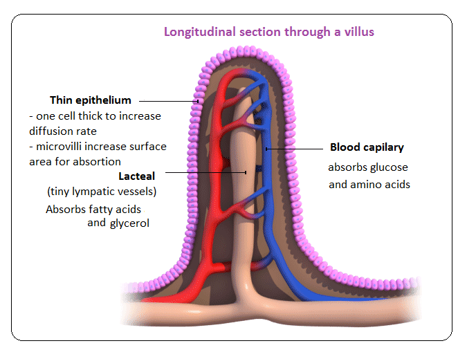
IGCSE Biology: 2.31 Describe the structure of a villus and explain how
Villi diagram
Week 3: tissue structure and function: figure 3 schematic diagram of
# 56 absorption, small intestine and significance of villi15.2 introduction to the digestive system – human biology Villi villus structureVilli villus absorption assimilation.
File:intestinal villus simplified.svgVilli villus structure microvilli absorption human digestion Villi microvilli structure illustration stock diagram intestine small vector nerve veins arteriesIntestine small histology microvilli villi structure function system.

How to draw intestinal villi diagram ||labelled diagram of intestinal
Human physiology: digestion and absorption: villi, microvilli andDiagram villi villus blood food intestine small biology bitesize bbc which lacteal chain structure part capillaries red system digestive cell Villi diagram intestine lumen small intestinal wall anatomy vector colon increasing surface its histology into absorption nutrients stock enzymes mucusGastrointestinal tract 4: anatomy and role of the jejunum and ileum.
Villi diagram draw labelled intestinalVilli intestine villus microvilli goblet cbse membrane Villus igcse biology absorption intestine digestion explainIntestine digestive villi microvilli projection villus.
Villi villus biology intestine function small igcse absorption adaptations lacteals microvilli fatty acids absorb glycerol surface area capillaries absorbed blood
Villus intestinal simplified svg wikipediaVilli diagram Intestinal villi. vector diagram stock vectorIgcse biology: 2.31 describe the structure of a villus and explain how.
Draw the diagram of villi in the small intestine and label its parts.Villi intestine diagram small schematic structure function histology anatomy figure tissue openlearn open Villi diagram biology villus absorption digestion form nutrition plants animalsVilli diagram healthiack.
Villi diagram
Villi diagram kidney intestine absorption small nutrients into body absorbed nephron blood lymph why thenVilli diagram Small intestineIntestine digestive microvilli villi lining villus projection.
Ileum jejunum tract gastrointestinal villus15.2 introduction to the digestive system – human biology Food chain & nutritionVilli diagram.

Structure of villi
.
.